|
PSVue® is a robust probe for detecting apoptosis. The PSVue® product family of fluorescent imaging reagents is based upon the positively charged bis(zinc-dipicolylamine) (Zn-DPA) (Smith, Bradley; US Patent 7,179,616) that functions as an Annexin-V mimic by targeting anionic phospholipids such as phosphatidylserine (PS). Therefore, PSVue® binds to the surfaces of apoptotic and necrotic cells which contain exposed PS.
Figure 1 depicts a generalized structure and mechanism of action of PSVue® probes. The PSVue® probe consists of an organic scaffolding which coordinates two positively-charged Zn2+ ions to form a complex (the "affinity group"). The organic component is conjugated, via a chemical linker, to a reporter group which is a fluorescent moiety1. Zn2+ then induces strong association of the complex through electrostatic interactions with carboxylate and phosphate anions present in the PS headgroup of dead and dying cell membranes, in turn leading to fluorescent labeling of the target bearing exposed PS2. PSVue® has been found to be highly selective for phosphatidylserine on vesicle membranes, compared to other species of phospholipids
 Figure 1
Structure and mechanism of action of PSVue® fluorescent probes. (a) Schematic diagram of PSVue® probes (b) Mechanism of binding of PSVue® to cell surface containing anionic phospholipids (c) Change in fluorescence intensity I/I0 (ex 380, em 440 nm) of PSVue® 380 (1 mM) in HEPES buffer (10 mM, pH 7.2) upon addition of 100 nM unilamellar vesicles composed of POPC : POPS (50 : 50, filled circles), POPC : POPS (95 : 5, open circles), 100% POPC (squares). Smith et al., Israel J. Chem, 45, 20052 and Smith et al., Cell Death Differ., 10(12), 2003.
|
PSVue® for in vivo targeting and imaging of cell death
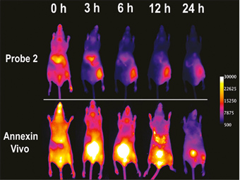 Figure 2
Representative NIR fluorescence images mice treated with cytoxic agent in the right leg and saline in the left leg injected with either near-infraed PSVue® 794 (top row-Probe 2) or Annexin-Vivo 750 (bottom row) via the tail vein 2 h post-treatment. (Smith et al., Mol. Pharm., 8(2), 2011
PSVue® for cancer imaging of (i) breast and (ii) prostate tumor
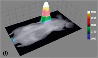 Figure 3 (i)
Representative overlay image of a nude mouse with an EMT-6 mammary tumor. Brightfield and fluorescence intensity images were acquired 24 h following injection of PSVue® 794 probe (ii) X-ray and fluorescence overlay image of a rat prostate tumor model at 24 h postinjection of PSVue® 794. (iii) Excised PAIII prostate tumors were sliced along the longest axis, and a 30 mm field of view generated the representative near-IR fluorescence intensity image. (Smith et al., J. Am. Chem. Soc., 132(1), 2010.)
|